ReProForce
FP7-REGPOT-2009-1
ReProForce logo
Slogun
REINFORCEMENT OF THE RESEARCH CAPACITY OF THE BULGARIAN INSTITUTE “BIOLOGY AND IMMUNOLOGY OF REPRODUCTION”
Events
- Jun 21, 2013
Information about the final meeting of ReProForce project participants /17-18 May 2013/
- May 10, 2013
Final meeting of ReProForce project
- Feb 28, 2013
Workshops of the ReProForce experts with business and scientific stakeholders in the IBIR-BAS
- Jan 30, 2013
Information for Open Doors Days in IBIR, BAS, November 29th – 30th 2012.
- Nov 14, 2012
Open Doors days – 2012
Mobility visit to the partner center Wageningen University, Human and Animal physiology group in Wageningen, Netherlands
Jul 16, 2012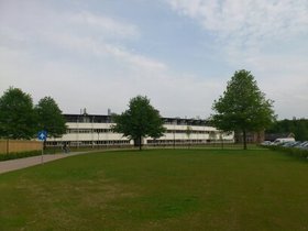
Participant: Elena Stoyanova, PhD student
The name of the visited institution: Human and Animal Physiology Group, Department of Animal Sciences, Wageningen University, Wageningen, Netherlands
Duration: 01.06.2012 - 30.06.2012
Purpose of the visit: Training of Histology and Microarray Analysis.
The research program included:
• Introduction to the research topics of the team and the used laboratory techniques. Main research topics are metabolism of nutrients, metabolic health and plasticity, effects of bioactive food components and physiological factors on the functions of different systems in mice.
• Visit of lectures:- High-content cell analysis – BD Pathway Brochure.
High Content Analysis (HCA) is the automation of fluorescence microscopy whereby the manual interpretation of images is replaced by computer algorithms that can quantify staining profiles in different areas of the image.
- Is sleep a waste of time?
Data was presented on the duration of the sleep in humans and different animals. A discussion concerning the topics followed:
Normal duration of sleep in humans.
Diseases affecting sleep duration.
Approaches for investigating and treatment of sleep disorders.
• Training for preparation of histology samples from mouse testis, adipose and muscle tissues. It demonstrated fixing and processing of tissues and organs (dehydratation, rinsing, infiltration and embedding) and cutting and staining of sections from tissues. Immunohistochemical staining for fast muscle myosin and polysaccharides was preformed.
Figure 1. Immunohistochemical staining of fast myosin, hematoxylin and eosin in mouse muscle fibers is shown on the Figure 1. The fast myosin is stained in brown, nuclei in blue and elastic, collagen and reticular fibers in pink.
Figure 2. Periodic-acid-schiff - hematoxylin staining in mouse muscle fibers. Polysaccharides are stained in dark violet; fibrin, plasma, collagen in bright violet and nuclei in blue. The polysaccharides staining (B) should be overlapping with fast myosin staining (A). The yellow stars mark the fibers that were stained according to the fast myosin staining (C).
• Training of microarray analyses techniques. It included isolation of total RNA, reverse transcription fluorescent incorporation, hybridization, scanning and analyses of results.
• Participation in laboratory seminars of the Human and Animal Physiology Group to present the results from my work in the lab and my current work on generation of induced pluripotent stem in IBIR.
• Visit to animal facilities for small and big animals of Wageningen University and introduction to the possibilities for performance of experiments with small animals.
Персонализирано търсенеМрежата
