ReProForce
FP7-REGPOT-2009-1
ReProForce logo
Slogun
REINFORCEMENT OF THE RESEARCH CAPACITY OF THE BULGARIAN INSTITUTE “BIOLOGY AND IMMUNOLOGY OF REPRODUCTION”
Events
- Jun 21, 2013
Information about the final meeting of ReProForce project participants /17-18 May 2013/
- May 10, 2013
Final meeting of ReProForce project
- Feb 28, 2013
Workshops of the ReProForce experts with business and scientific stakeholders in the IBIR-BAS
- Jan 30, 2013
Information for Open Doors Days in IBIR, BAS, November 29th – 30th 2012.
- Nov 14, 2012
Open Doors days – 2012
Report for mobility visit in partner center Jena, Germany
Jul 28, 2011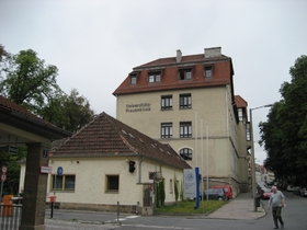
Participants: Georgi Georgiev, PhD student
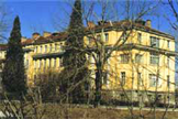
The name of the visited institution:
The host of the visit was Prof. Dr. Udo Markert, head of the Friedrich Schiller University’s Placenta Lab. The laboratory is located in Jena, Germany. Research in the Placenta Lab, part of the Department of Obstetrics, is focused on a variety of scientific topics in relation to pregnancy. They include immunological mechanisms of embryo implantation, regulation of trophoblast invasion, regulation of decidual immune cells as well as allergic, pharmacological and nutritional aspects. The Placenta Lab is member of the European Network of Excellence EMBIC ("The control of Embryo Implantation").
Duration: 14 June – 14 July 2011.
Purpose of the visit
The topic of the one-month project was “In vitro effects of glucocorticoids and melatonin on the expression profile of COV434 granulosa cell line”.
The research program included:
- Cultivation of COV434 granulosa cell line;
- In vitro experiments;
- Isolation of total RNA from cultivated cells according to specified protocol;
- cDNA synthesis (reverse transcription);
- End-point singleplex PCR;
- Agarose gel electrophoresis and visualization of cDNA products.
Week 1 (15 – 17 June):
The first week of the visit included introduction to the laboratory and development of detailed project plan and timetable. Along with the elaboration of the work protocols for the cell culturing and the PCR, the necessary chemicals and primers for the experiment were ordered. The visitor was introduced to cell culturing procedures and manipulations with the cells to be investigated. PCR was performed by employment of cDNA isolated from cells cultivated under normal conditions, subsequently analyzed on agarose gel electrophoresis according to the protocols of the lab. The first week included also introduction to some of the other methods used in the lab (FACS, single-cell PCR, etc.). The project, as well as the home institute (IBIR-BAS) was presented at the host laboratory meeting.
Week 2 (20 – 24 June)
The second week was assigned for conducting experiments with the granulose cell line. Total RNA from frozen cells was isolated according to the manufacturer’s protocol (innuPREP RNA mini kit, Jena Analytik), followed by cDNA synthesis using GoScript™ Reverse Transcription System kit. The synthesized cDNA was then used for testing of 9 primers chosen to test the functional capacity of the cultivated granulose cells.
Week 3 (27 June – 01 July)
During the third week of the visit the experimental cultivation of the cells was accomplished. The cells were collected for PCR analysis, and the supernatants – for measurement of steroids. PCR with template cDNA and genomic DNA was performed, followed by agarose gel electrophoresis and UV – imaging. The visitor was invited to attend seminar on “qRT-PCR”.
Week 4 & 5 (04 – 08 July & 11 – 13 July)
Total RNA was isolated from all experimental and control samples using the above described kit protocol. RNA concentration was measured with Nanodrop™, and the results confirmed the purity of the isolated material. The total RNA was then used for reverse transcription using kit protocol (GoScript Reverse Transcription System). Each of the samples was used for cDNA synthesis along with NTC (no template) control and no reverse transcriptase control (RT-). Each of the cDNAs and control samples was then used for PCR with the 9 primers specified above. Total of 324 PCR samples were run and then used for agarose gel electrophoresis. 12 x 2% agarose gels were run and imaged using UV according to the protocols used in the lab. The results were presented during a lab meeting on 12 July 2011.
Персонализирано търсенеМрежата
IBIR-BAS
-
Links
-
-
Committee
-