ReProForce
FP7-REGPOT-2009-1
ReProForce logo
Slogun
REINFORCEMENT OF THE RESEARCH CAPACITY OF THE BULGARIAN INSTITUTE “BIOLOGY AND IMMUNOLOGY OF REPRODUCTION”
Events
- Jun 21, 2013
Information about the final meeting of ReProForce project participants /17-18 May 2013/
- May 10, 2013
Final meeting of ReProForce project
- Feb 28, 2013
Workshops of the ReProForce experts with business and scientific stakeholders in the IBIR-BAS
- Jan 30, 2013
Information for Open Doors Days in IBIR, BAS, November 29th – 30th 2012.
- Nov 14, 2012
Open Doors days – 2012
Report for mobility visit in partner center Jena, Germany
Apr 3, 2012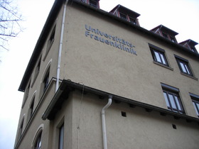
Participant from IBIR: Milena Mourdjeva
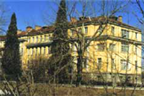
The name of the visited institution: Placenta Labor, research group of the Department of Obstetrics, Universitatsfrauenklinik, Friedrich Schiller Universitat, Jena, Germany (http://www2.uni-jena.de/ufk/placentalabor/cms/).
Head of the Placenta Labor is Prof. Dr. Udo Markert, ReProForce project partner.
The Placenta Lab, the research group of the Department of Obstetrics, is focused on a variety of scientific topics around pregnancy. Those include immunological mechanisms of embryo implantation, regulation of trophoblast invasion, regulation of decidual immune cells as well as allergological, pharmacological and nutritional aspects. Respectively, the specializations of students performing their diploma or doctoral theses are multifold. Our students derive from faculties of medicine, biology, biochemistry, chemistry and nutritional sciences.
The Placenta Lab is member of the European Network of Excellence EMBIC ("The control of Embryo Implantation")
Duration: 16 January – 15 February 2012.
Purpose of the visit:
The topic of the one-month project was “Progesterone Induced Blocking Factor (PIBF) expression in Decidual Mesenchymal Stem Cells (DSC)”.
The research program included:
- Immunoblot evaluation of PIBF expression in Mesenchymal Stem Cell (MSC) isolated from bone marrow (BM-MSC), adipose tissue (ASC) and early decidua (DSC);
- PCR for mRNA- PIBF expression in Mesenchymal Stem Cell (MSC) isolated from bone marrow (BM-MSC), adipose tissue (ASC) and early decidua (DSC);
- siRNA silencing of PIBF in primary cell cultures;
- Isolation of Placental Mesenchymal Stem Cell (pMSC)
The visit started with introduction to the laboratory and development of detailed project plan. The visit included also introduction to some of the methods used in the lab (migration test, invasion test, etc.). The project, as well as the home institute (IBIR-BAS) was presented at the Placenta lab meeting on the last day of the visit. Results obtained by Milena Mourdjeva during the visit in the Placenta Lab were discussed as well as future perspectives for the investigations on the role of PIBF in early pregnancy. The opportunity to participate in the weekly lab meetings (each Tuesday morning) and Journal club (each Wednesday evening) was given to Milena. One of the Wednesday meetings was dedicated to the Sigma presentation about Zinc Finger Nuclease Technology – a method for targeted editing of the genome by creating double-strand breaks in DNA at user-specified locations.
- Immunoblot evaluation of PIBF expression in Mesenchymal Stem Cell (MSC), isolated from bone marrow (BM-MSC), adipose tissue (ASC) and early decidua (DSC)
Proteins obtained from Trizol fractions of cells after RNA and DNA isolation were determined by Bradford method. The amounts of protein for each cell line were equilibrated. Electrophoresis, transfer and immunoblot were performed according to protocols used in Placenta Lab. Protocols and buffers used in Placenta Lab are useful upgrade to the methodology, used in IBIR.
Results: All three types of mesenchymal stem cells tested showed significant amount of PIBF detected by rabbit polyclonal antibody. Future experiments including the 3A6 anti-rhPIBF monoclonal antibody can specify and confirm the PIBF splicing variants expressed by mesenchymal stem cells.
- PCR for mRNA - PIBF expression in Mesenchymal Stem Cell (MSC), isolated from bone marrow (BM-MSC), adipose tissue (ASC) and early decidua (DSC)
PCR was performed with cDNA isolated from BM-MSC, ASC and DSC and subsequently analyzed on agarose gel electrophoresis according to the protocols of the Placenta lab. Total RNA from Trizol treated cells was isolated, followed by cDNA synthesis using GoScript™ Reverse Transcription System kit. The synthesized cDNA was then used for testing of 3 primers sets for PIBF. Agarose gel electrophoresis and UV – imaging were applied to visualize the PCR products. The principles of programs for primer design and primers evaluation were discussed.
Results: One of the tree primer pairs tested is suitable for PIBF mRNA evaluation. Conditions for PCR reactions were adjusted. The work on PIBF mRNA expression levels in DSC will be continued in IBIR.
- siRNA silencing of PIBF in primary cell cultures
Placenta lab protocols for transfection of cells with PIBF siRNA were applied to primary culture mesenchymal stem cells to silence the PIBF protein expression. PIBF siRNA #1 and #3 were used for MSC transfection with TurboFect (Fermentas) for 24 (only #1) and 72 hours. After incubation, the cells were lysed with Lysis Buffer (Cell Signaling), protein amounts were determined with Bradford reagent (BioRad), equal protein amount of siRNA treated and control cells were loaded for electrophoresis and blotted with anti-49kDa rhuPIBF rabbit IgG (RabbitN1, II bleeding; protein G purified rabbit IgG). The blots were developed with Luminate Forte (Millipore) and the membrane was stripped with mild stripping buffer (Abcam) and incubated with anti-bActin antibody as loading control antibody. The reaction was developed again using the same revelation method and bands for PIBF and bActin were analyzed.
Results: There were no differences in PIBF expression levels between treated and control cells between any of the tested conditions. Either the applied siRNA or the used protocols were not suitable for PIBF silencing in MSC.
- Isolation of Placental Mesenchymal Stem Cell (pMSC)
On the request of the hosts, the applied protocol for isolation of decidual mesenchymal cells at the visitor’s lab was used for isolation of term placenta mesenchymal stem cells. Tissue from placenta was cut into small pieces, and incubated with collagenase. The obtained cell suspension was centrifuged, the cell pellet was resuspended in DMEM (PAA) and the cells were seeded. The media was changed after 24 h and thereafter every other day.
Results: The protocol needs some additional optimization (usage of DNAase and cell strainer) but the obtained cells possess the morphological features of mesenchymal stem cells. On the day after isolation the single fibroblast like cells were attached to the culture dish. The cells proliferate and form the confluent monolayer up to day 10.
Персонализирано търсенеМрежата
